Civil War Hospital Ship

The U.S.S. Red Rover, a captured Confederate vessel, was refitted as a hospital ship.

Evolution of Civil War Nursing

The evolution of the nursing profession in America was accelerated by the Civil War.

The Practice of Surgery

Amputations were the most common surgery performed during the Civil War.

Army Medical Museum and Library

Surgeon-General William Hammond established The Army Medical Museum in 1862. It was the first federal medical research facility.

Civil War Amputation Kit

Many Civil War surgical instruments had handles of bone, wood or ivory. They were never sterilized.

Showing posts with label Civil War Medical Instruments and Supplies. Show all posts

Thursday, March 2, 2017

Porcher’s Resources of the Southern Fields and Forests

From: waring.library.musc.edu

It is intended as a repertory of scientific and popular knowledge as regards the medicinal, economical, and useful properties of the trees, plants, and shrubs found within the limits of the Confederate States, whether employed in the arts, for manufacturing purposes, or in domestic economy, to supply a present as well as a future want.
Francis Peyre Porcher
Preface, page iii

At the outbreak of the Civil War, Francis Peyre Porcher joined the Confederate Army as a surgeon to South Carolina’s Holcombe Legion and was then transferred to the Naval Hospital at Norfolk, Virginia in March 1862. He finished his Confederate service in the South Carolina Hospital at Petersburg, Virginia. While in Virginia Porcher was “released temporarily from service in the field and hospital” [Preface, page iii], by C.S.A Surgeon General Samuel Preston Moore to write, Resources of the Southern Fields and Forests, Medical, Economical and Agricultural; Being Also a Medical Botany of the Confederate States; with Practical Information of the Useful Properties of Trees, Plants, and Shrubs, first published in Charleston in 1863. The handbook identified local plants with therapeutic qualities that could be used not only by Confederate surgeons, but planters and farmers, in place of manufactured drugs made unavailable because of the Union blockade. The handbook was of such value that a revised edition was published in 1869.

The Regimental Surgeon in the field, the Physician in his private practice, or the Planter on his estate may themselves collect and apply these substances within their reach …
Preface, page iii

Four editions of the manual were produced during the course of the war and are now available for research use at the Waring Historical Library:
1863 edition
1869 edition
1863 edition, [transcription]

1869 edition
I here introduce a notice of upwards of four hundred substances, possessing every variety of useful quality. Some will be rejected as useless, others may be found upon closer examination to be still more valuable. The most precious of all Textile Fibres, and Grains, Silks, Seeds, Oils, Gums, Caoutchouc, Resins, Dyes, Fecula, Albumen, Sugar, Vegetable Acids, Starch, Liquors, Spirit, Burning Fluid, material for making Paper and Cordage, Barks, Medicines, Wood for Tanning and the production of Chemical Agencies, for Timber, Ship-building, Engraving, Furniture, Implements and Utensils of every description--all abound in the greatest munificence, and need but the arm of the authorities or the energy and enterprise of the private citizen to be made sources of utility, profit, or beauty.
Preface, page vii-viii

Wednesday, January 18, 2017

Advancements in Surgery Through the Ages

by Lisa J. Fulghum, 6-5-15

Imagine yourself standing in a medical tent on September 17, 1862 not far from Sharpsburg, Maryland, as the bloody battle of Antietam rages on the other side of the bridge. With nearly 22,000 dead and many more wounded after just 12 hours of fighting, what is a good surgeon to do? Your shirt is stained with the blood of the many you have already treated, yet more men than you can count lay suffering nearby. With only 98 medical officers to serve the entire Union army and just 24 for the Confederates when the war began, trained medical professionals are still hard to find.1 It will be a very long night. Your options are as limited as the precious time you have.

Ether and chloroform (when they are available) are the only viable methods of anesthesia, besides a stiff shot of whiskey. The best surgeons, commonly called butchers by reporters and patients, are the ones who are quick and hopefully accurate. Amputation is the most common surgery of the day, and a good surgeon can amputate a limb in just 15 minutes.2

Standing amidst the grizzly scene, you call for another cup of coffee, wipe your surgical saw on your apron and look for the nearest patient who is likely to survive long enough to complete a surgery. Inwardly, you hope that one day you will have a better method for dealing with such carnage.

An estimated 60,000 amputations were performed during the Civil War. Nearly 75% of amputees survived the surgery, though that depended greatly on the limb that was amputated. A forearm amputation mortality rate was 14%, but legs amputated at the hip had an 88% mortality rate.3

History of Surgery
Since the beginning of time, man has developed and refined instruments for surgical procedures and for healing people. Throughout our history, humankind has had notable physicians who made monumental contributions to medical science.

As early as 600 B.C. Sushruta , India’s preeminent surgeon, pioneered rhinoplasty and other forms of plastic surgery. Since cutting off a person’s nose was a common punishment, Sushruta’s expertise was highly valued.4 Among other things, believe it or not, he even documented cataract surgery.5 Developing and documenting 120 surgical instruments (in bronze, silver, and iron) and employing many relatively modern techniques, Sushruta was revered as a master physician 150 years before Hippocrates.

Around 1000 A.D. Abu al-Qasim al-Zahrawi (a.k.a. Albucasis in Europe), the master Muslim physician of Spain, wrote a 30-volume medical encyclopedia called Al-Tasrif (The Method of Medicine). It was the medical reference text used for over 500 years. Albucasis invented and described over 200 new instruments. With hand sketches of what instruments looked like, how they were constructed and how they were to be used, he documented instruments like his obstetric forceps and an urethrascope. Not only was he the first author to describe an ectopic pregnancy and the hereditary nature of hemophilia, but he also pioneered sublimation and distillation for the preparation of medicines. Albucasis described ligatures 600 years before Paré adopted their use, even using catgut for internal stitches so the stitches would dissolve as the patient healed.6

Surgery was not always the prestigious profession that it is today. During the middle ages in Europe, surgery was considered a lesser profession, and physicians did not deal with such things. The local barber was frequently the town surgeon.

Stop the Bleeding
During the Middle Ages, one of the major hindrances of a successful surgery was excessive bleeding. Medical innovators like Guy de Chauliac emerged. He released (1363) his 7-volume library of surgical knowledge, the Chirurgia Magna, in which he described the use of a tourniquet for blood control during an amputation. In his work, he also described intubation, suturing, blood clotting, and the use of surgical instruments.7

By the mid-1500s Ambroise Paré, a barber-surgeon, introduced the lost art of ligatures to control blood flow in wounds or during surgeries like amputation. He favored ligatures over cauterization that was the preferred method of the day, because ligatures reduced the pain patients suffered. Paré invented the crow’s beak hemostat to stop blood flow during amputation.8

Kill the Pain
The pain of surgery was a limiting factor. A doctor could only operate for the length of time a patient could endure. The introduction of anesthesia opened the door for a host of new procedures.

Humphrey Davy, the scientist who discovered the elements potassium and sodium, was one of the first to identify the anesthetic properties of nitrous oxide and ether in 1799. Both were used as recreational drugs at the time. In 1842 Dr. Crawford W. Long, who had observed the effects of ether at some of their nitrous oxide parties, was likely the first to use it in surgery. He administered it to James Venable before removing a cyst from James’ neck.9

In Scotland a year later (1847) James Young Simpson was assisting a woman with a difficult childbirth and used ether to minimize her pain. Because of the side effects, he began experimenting with chloroform. Chloroform was soon accepted, and even Queen Victoria used it for childbirth.

Physicians then began looking for a local anesthetic. The birth of the hypodermic needle (1850s) allowed for intravenous anesthetics.10 Surgeons like William Stewart Halsted, who was one of the “Big Four” founding professors of Johns Hopkins, was one of the first to explore the use of cocaine as a local anesthetic. He would inject the anesthetic into a nerve trunk to numb an entire limb or the spinal cord.11

Incidentally, it was he who introduced the radical mastectomy for treatment of breast cancer. In addition, he also invented the Halsted Mosquito Forceps, a small pair of hemostatic forceps. One of the other “Big Four” was Howard Atwood Kelly, who developed the Kelly Forceps, one of the most popular and commonly used hemostatic forceps.

With the introduction of anesthetics, medical science continued its forward advance. However, the discovery of effective anesthetics came with unintended consequences. While it made non-traumatic surgery possible and allowed for longer and more complicated surgeries, it also opened the patient to a greater chance of infection.12 By the mid-1800s when the use of anesthetics was the norm, it was not uncommon for a European hospital to have an 80% mortality rate for surgical patients.13

Wash Up
When physicians learned to combat infection, we cleared another medical hurdle. Honey, willow bark, balsam, wine, and vinegar were used historically as antiseptics to dressed wounds. However, before the mid-1800s, there was little concern for cleanliness among physicians. At that time, a bloody apron was considered a badge of honor. In 1795, Scottish physician Alexander Gordon suggested that doctors could transmit contagions, and he recommended that obstetricians wash their hands and clothes before treating patients. Opposing the standards of the day, American physician Oliver Wendell Holmes concluded in 1843 that the spread of puerperal fever (after childbirth) was connected with unwashed doctors. Independently, Hungarian doctor Ignāz Semmelweis made the same conclusion in 1847. Implementation of new cleanliness guidelines lead to a maternal mortality drop from a high of 18% to about 1%. Amazingly, these doctors understood what to do but not why.14

Louis Pasteur proved the existence of airborne microorganisms. By 1862, he completed his first tests indicating that bacteria cause wine, beer, and milk to sour. He then pioneered the process of pasteurization to kill the microorganism. Pasteur continued his extraordinary research in germ theory and went on to invent vaccinations for diseases like cholera, tuberculosis, and small pox.15

Following these discoveries, Joseph Lister put it all together. He determined that if airborne bacteria caused wine to spoil, then airborne microorganisms could also cause infection after surgery or childbirth. In 1865, he pioneered a method of using carbolic acid (phenol) as an antiseptic for treating wounds. He published his work in 1867 in The Lancet. It was not fully accepted until the 1890s when Heinrich Koch proved that germs cause disease.

Lister introduced sterile catgut for internal stitches. He also tied bones together with sterile, silver wire and left it inside of the patients. This was a revolutionary idea, because previously, the germs left behind by such a procedure would have caused gangrene and certain death.16 Lister also invented the Lister bandage scissors.

Hurdles to Modern Surgery
While the mechanics and instruments in a surgeon’s toolkit have changed dramatically through the ages, surgery still remains an intricate and delicate handicraft practiced and perfected by the gifted and skilled. As the science of surgery has evolved, three major hurdles had to be overcome: the control of bleeding, the control of pain, and the control of infection. In the last 150 years, these incredible medical advancements have completely revolutionized the surgical process and given humankind a much brighter future.

Now, as you stand in a modern operating theater, garbed in a sterile gown complete with surgical mask, binocular loupes and neoprene gloves, you can marvel at the array of gadgetry to monitor the patient’s heart rate, O2 levels, and blood pressure. The lead surgeon is assisted by a staff of anesthesiologists, nurses, and technicians. Complex surgeries of critical organs can take hours as skilled surgeons work miracles never before even imagined. The civil war surgeon would have marveled at the spectacle of a laser surgery, laparoscopy, MRI and CT technology, chemotherapy, and robotic surgery. Given the phenomenal advances of the last century, we have to wonder what revolutionary breakthroughs await us in the next 150 years. Will you be the Louis Pasteur, Ambroise Paré, Albucasis, or Joseph Lister of our modern age?

References

Civil War Medical Care, Battle Wounds, and Disease (Civil War Medical Care, Battle Wounds, and Disease) www.civilwarhome.com/civilwarmedicine.html 02/10/02
Clements, I.P. History of Surgical Amputation (HowStuffWorks) health.howstuffworks.com/medicine/modern-treatments/amputation2.htm
Billings, J. (2001). Hardtack and coffee, or the unwritten story of Army life. Scituate, Mass.: Digital Scanning. www.civilwar.org/education/pdfs/civil-was-curriculum-medicine.pdf
ISPUB.com (Internet Scientific Publications) Sushruta: The first Plastic Surgeon in 600 B.C. by S Saraf, R Parihar ispub.com/IJPS/4/2/8232
Roy, P., Mehra, K., & Deshpande, P. (1975). Cataract surgery performed before 800 B.C. British Journal of Ophthalmology, 59(171) bjo.bmj.com/content/59/3/171
Albucasis (Doctor - the Father of Modern Surgery) english.turismodecordoba.org/seccion/albucasis-doctor---the-father-of-modern-surgery
Guy de Chauliac www.thefamouspeople.com/profiles/guy-de-chauliac-447.php
Ambroise Paré www.apimsf.org/default.aspx?id=22
Dr. Long’s Discovery www.crawfordlong.org/id10.html
Blatner, A. (2009, February 16). Anesthesia / History of Medicine. The Discovery and Invention of Anesthesia. www.blatner.com/adam/consctransf/historyofmedicine/4-anesthesia/hxanesthes.html
The Four Founding Physicians http://www.hopkinsmedicine.org/about/history/history5.html
Antisepsis www.discoveriesinmedicine.com/A-An/Antisepsis.html
Lamont, A. (1992, March 1). Joseph Lister: Father of Modern Surgery. https://answersingenesis.org/creation-scientists/joseph-lister-father-of-modern-surgery/
Henderson, D.K., Lee, L., and Palmore, T (2014, June 1) The Contemporary Semmelweis Reflex: History as an Imperfect Educator www.infectioncontroltoday.com/Articles/2014/06/The-Contemporary-Semmelweis-Reflex-History-as-an-Imperfect-Educator.aspx
Louis Pasteur (2014) www.biography.com/people/louis-pasteur-9434402
Antisepsis

A Purdue University graduate, Lisa Fulghum is the Content Director at WPI. For nearly 50 years, WPI has been supplying laboratory equipment for life science researchers, including a variety of German and Swiss made surgical instruments.

Image: This toolkit is over 100 years old.

From: alnmag.com

Tuesday, November 29, 2016

Amputations

From: fisher.k12.il.us

Amputations were a common surgery performed in the civil war. Doctors said that it saved many more lives than it killed. Survival rate for amputations done in the first 24hrs. After an injury was very good with mortality. If they were done after the first 24hrs -mortality rate doubled to 50%. Surgeons tried to go as fast as possible. It’s generally a myth that most operations were performed without anesthesia with only a bullet to bite.

Their procedure:

1st-they cut off blood flow with a tourniquet.

2nd-after that he’d take a scalpel and slice through the outlying tissue and flesh.

3rd-Then he’d use a hacksaw-like tool called a capital saw to saw through the bone. (It had replaceable blades.)

4th-after bone and flesh was all sliced off the surgeon would take silk sutures in the north and cotton sutures in the south and sew the major arteries and veins together. This limb would be dropped on a pile that got thrown out on the platform. This would take about 15 min.

Amputation tools

Proving an amputation or surgical kit was used in the Civil War is difficult. There were two types of amputation blades, The circular blade which would more than likely be earlier then the Civil War, and the straight blade about the time of the Civil War.

Wednesday, June 29, 2016

History Affects Morphine: The Hypodermic Needle

From: itech.dickinson.edu, 4-30-08

“Ah! Pierce me one hundred times with your needle fine
And I will thank you one hundred times, Saint Morphine,
You who Aesculapus has made a God.”
- Jules Verne
(Poem taken from In the Arms of Morpheus by Barbara Hodgson)

Despite its impact on the science of pharmacology, morphine had limited medical impact until the invention of the hypodermic needle in the 1840s/1850s

A number of individuals are associated with the invention of the hypodermic needle, but among them, Alexander Wood, a Scottish physician, is perhaps the most prominent

Wood used morphine in conjunction with his newly invented needle to treat a patient with neuralgia, otherwise known as a sharp pain in the nerves; unfortunately, Wood also used his device on both he and his wife, and both became addicted. In fact, Wood’s wife became the first woman to die of a narcotic drug overdose

In light of this, however, Wood found that upon injection, morphine’s results were both immediate and much more powerful, certainly a success; such success led to a rise in the medical use of morphine, especially in the realm of surgery and anesthesia

Unfortunately, though, morphine administered through hypodermic needle was not thought to be addictive, and thus it further proliferated the drug, increasing use and addiction

Interestingly, it is worth noting that shortly after the time of the hypodermic needle’s inception, there began a debate over whether the effects of morphine post-injection were localized or not; and while many believe the effects to be non-localized – hence the name hypodermic – the debate actually continues to this day

Image: historical perspective: old syringes


Tuesday, May 24, 2016

Civil War Pharmacy: A History of Drugs, Drug Supply and Provision, and Therapeutics for the Union and Confederacy (review)

Reviewed by Gregory J. Higby

Michael A. Flannery. Civil War Pharmacy: A History of Drugs, Drug Supply and Provision, and Therapeutics for the Union and Confederacy. New York: Pharmaceutical Products Press (Haworth Press), 2004. xiii + 358 pp. Ill. $59.95 (cloth, 0-7890-1501-3); $34.95 (paperbound, 0-7890-1502-1).

In lieu of an abstract, here is a brief excerpt of the content:
As is often the case today, a work's subtitle tells the full story of its contents. While Michael Flannery gives attention to the traditional aspects of pharmacy—the making of medicines—he focuses on the drugs themselves and their use. Forty years ago, when George Winston Smith wrote Medicines for the Union Army, history of pharmacy, like the discipline it studied, was limited to the production and distribution of medicines. Today the field of pharmacy includes pharmacology, therapeutics, and drug utilization, an expansion reflected in Civil War Pharmacy. Flannery has written a valuable book for both buffs and historians. For the lover of Civil War lore, the text yields useful details and captivating stories. For the professional, the author provides a solid narrative, cogent interpretation, and good documentation. Not one to shy away from controversy, Flannery tackles a few "old chestnuts," including the court martial of Surgeon General Hammond and the debatable efficacy of Civil War therapeutics.

The narrative of Civil War Pharmacy is divided into three parts, followed by a set of exceptionally useful appendices that include the Union and Confederate standard supply tables for medicines, a long excerpt from The Hospital Steward's Manual, and the text of Hammond's famous Circular No. 6. In part 1, Flannery "sets the stage" with three chapters—one largely historiographic, another on the "State of Pharmacy in America, 1861," and a third examining women and Civil War pharmacy. Readers knowledgeable about Civil War medicine should resist the temptation to skip the first chapter, because mixed in with the expected references to Doctors in Blue and laudable pus is a solid argument for the importance of medicines in the conflict: because of the high rate of treated diseases—malaria, respiratory ailments, and digestive disorders—"the medical context for the Civil War . . . was not one principally of saws and sutures but of mortar and pestle" (p. 23).
In part 2, Flannery concentrates on pharmacy in the Union with chapters on purveyors, manufacturing, and therapeutics. This is the strongest section of the book. An entire chapter is devoted to calomel and quinine, two favorites of allopathic physicians of both armies. While the calomel story, wrapped up with the downfall of William Hammond, might be more sensational, Flannery's discussion of quinine as drug and commodity is enlightening. After this, part 3 on the Confederacy is a bit of a disappointment. Flannery bears little blame for this: Many of the records of the South were lost. More importantly, there was much less pharmacy in the South—almost no significant medicine manufacturing existed there before the war, and the Union's naval blockade hampered the importation of drugs. (The vast majority of botanical drugs came from abroad.) Instead, Flannery's story shifts from laboratories and tons of output in the North to the South's blockade runners and the valiant attempt of Southerners to find substitutes for foreign drugs. [End Page 774]

Flannery concludes his book with a short epilogue arguing persuasively that the Civil War had sweeping influences on the development of the American pharmaceutical trade in the late 1800s. Forced by an incredible increase in demand, companies like Squibb, Wyeth, and Sharp and Dohme industrialized their operations—ending the era when the drug business was a cottage industry.
Because this is a work on the Civil War, its nearly inevitable popularity should elicit a second edition. If so, I encourage the author to consider the following suggestions: First, insist on better production values from your publisher: the uninteresting typography and murky photographic reproduction detract from the work; also, add a few maps showing laboratory locations and other sites of interest. Second, avoid "pharmaceutical care," a loaded term that carries a specific meaning for today's pharmacists that is not equivalent to "medical care." Third, carefully go over the quinine/cinchona sections: in some places, cinchona is called a derivative of quinine...

From: Bulletin of the History of Medicine
Volume 80, Number 4, Winter 2006
pp. 774-775 | 10.1353/bhm.2006.0129

From: muse.jhu.edu

Monday, April 11, 2016

Bandages and Adhesive Plaster

By Glenna R. Schroeder-Lein

Caring for wounds during the Civil War usually involved the use of dressings and bandages of some sort. At the first aid station an attendant would apply a bandage to stop the bleeding. This could be a rolled bandage of clean cotton or linen from a well-stocked medicine chest or wagon. Or, during a desperate battle with crowds of patients exhausting the supplies, the bandage could be a strip of the doctor's shirt.

Once the wound had been cleaned and any necessary operation performed, the wound was often covered with cerate (a beeswax ointment) and then a dressing made of lint (fibers scraped from old linen cloth) or raw cotton (sometimes baked until charred). Most doctors used a cold-water dressing, keeping the pad moist with cold water through frequent applications or a continual drip. Some used warm dressings, but these usually produced infection ("laudable pus"). On occasion some doctors used dry dressings with successful healing and fewer infections.

Dressings were held on the wounds with bandages. Many women, North an South, gave up sheets, bedspreads, skirts, and other material to be torn into strips and rolled for bandages, which were used in addition to manufactured bandages. While these bandages were clean after a fashion, they were never sterile. In many hospitals, bandages were washed and reused, often because of shortages, sometimes spreading infections. In other hospitals, doctors refused to ruse bandages because they suspected that reused bandages hindered healing. Although not elastic, bandages could also be used in the manner of modern elastic bandages to affix splits to broken limbs.

Civil War doctors also used "adhesive plaster." This sticky bandage was made by spreading a mixture of resin, olive oil, lard, and lead monoxide on this muslin. The dried bandage was then rolled for storage and transportation. It could be cut in strips of any length and width for a variety of uses, such as holding the edges of a wound together, holding dressings and bandages in place, and helping to stabilize fractures. However, adhesive plaster bandages could not simply be stuck on; they had to be heated to make them stay. Many doctors kept a can of hot water handy and put the back of the adhesive strip around it until it was sticky enough for use. The bandages could also be heated over the flame of a spirit lamp. If all else failed, rubbing the bandage between the thumb and forefinger might warm the bandage enough for use. Because of the problems of heating the adhesive plaster bandages and because they sometimes irritated the patient's skin, not all doctors used them.

Plaster bandages, such as are used to make casts, were uncommon and were not a regular part of the hospital supplies.

From: "The Encyclopedia of Civil War Medicine" by Glenna R. Schroeder-Lein

Wednesday, February 24, 2016

A Historical Glimpse of Suturing

From: theapprenticedoctor.com

Suturing is commonly used today as a means by which wounds are closed in order to prevent infection and hasten healing. Stitches are used by surgeons, doctors, nurses, podiatrists, dentists, first aid workers, veterinarians and survivalists. However, where did the concept for suturing wounds begin?

Archeologists have discovered primitive needles of bone and later metal that were thought to be used for surgical suturing thousands of years ago. It is suggested that the original versions of suture threads were coarse and made from plant fibers before progressing to animal parts much later in history. The first obvious proof of using sutures surgically comes from the Egyptians who used the technique extensively for burial preparation of their mummies. It is assumed that they also used their suturing skills on living people as well. The Greek physician Hippocrates recorded his use of sutures and plant-based materials, but the crude methods and materials most assuredly led to infections and severe scarring during that time of use.

Although the exact date of the transition from plant fibers to animal suturing materials is unknown, the first recorded use of catgut as a suturing material is attributed to Galen of Pergamon in the second century AD. Catgut was and still is made from cattle and sheep intestines, although synthetic catgut is more commonly used today, but like its predecessor, does not require removal because it dissolves in the body.

One of the main problems with suturing surgical incisions and wounds early on was that sterile instruments were not used which often led to skin irritations and infections. In 1860, Joseph Lister addressed the problem by creating a method of for sterilizing suture material by using carbolic acid. Chromic acid was later used to achieve sterilization and then iodine was introduced to sterilize catgut in 1902. These substances greatly decreased the incidents of infection in sutured wounds.

Sterilized catgut has been successfully used for decades and is still in use today. Eventually, however, doctors began to seek other suture options because catgut dissolved unpredictably in different patients which sometimes led to wounds reopening. Scarring was also a major problem with catgut sutures. Synthetic suture materials were produced in the 1920’s and the 1930s saw the creation of two types of surgical sutures, absorbable and non-absorbable which offered doctors a broader choice in suturing materials.

Today, most suture kits contain suture thread made from polymer fiber materials which work well for a variety of surgical suturing procedures. Polymer fiber suture thread comes in a wide selection of styles, designs and types which can be utilized for various wounds and under numerous conditions. Due to its flexible nature, polymer fiber is the preferred choice of physicians, nurses, dentists, veterinarians and first responders and is the best selection for survivalists, adventurers, sports teams and high-risk job workers. Suturing has come a long way since its conception and suture kits offered today contain top sterile equipment and materials to perform wound stitching properly which significantly reduces both infection and scarring.

Suture Pins

By Dr. Michael Echols, 2-4-12

Pins used to approximate the sides of a soft tissue incision, from an 1864 Tiemann surgery set.  The pins are round with a round, not a flat head.  These needles were found in the paper holder and would have been supplied this way.

In the diagram, the pins are shown with suture material wrapped  in a criss-cross on either side of the incision and tied to close the incision, rather than being passed through the tissue.

An image of a Buck's suture pin director is shown, which would have been used to guide the placement of a pin.

From: medicalantiques.com

Monday, February 1, 2016

Civil War Sutures

Sources: Rita, Oconto County Coordinator, WiGenWeb  and Joan Benner, Adams and Marquette County WiGenWeb Coordinator

After the war, 1861 - 1865, the medical doctors from North and South came together in an attempt to exchange what they had learned. This information was to be used in the emergency treatment of civilians.

One glaring difference was the substantially higher number of ancestors who survived major surgery in the South than in the North, where the post treatment infection rate caused high numbers of
fatalities.

The doctors of the North (often called "sawbones" for all the amputations done) used imported silk thread for stitching wounds. It was strong, light and "slick" , making use much easier during
stitching.

Because of the naval blockades, there was no silk thread in the South, for even the most prominent of patients. Cotton thread was not a success as it broke down before the wound had healed
sufficiently. Horse hair was tried. But because it was too stiff to make sutures in it's natural state, it was boiled in water to soften and become more pliable.

The result was that the Northern silk thread had been handled by countless unwashed hands from several continents and introduced all the pathogens it carried right into the wound.

The horse hair was sterile when it came from boiling, and handled by significantly fewer numbers of people. Far fewer pathogens were introduced right into those wounds.

With antibiotics and sterile procedural practices still years away, many folks owe their ancestral branches to the boiled horse hair sutures, and to the women and men who devised that method of
softening the fibers for using in textiles.

Rita - Oconto County WIGenWeb Coordinator

From: wiroots.org

The History of the Hypodermic Needle

By Nick Snelling, 7-15-14

Who do we have to thank for the invention of the squeamish yet fantastically clever hypodermic needle? Health humorist Nick Snelling finds out...

Whether it’s an anaesthetic, blood test, insulin, vitamin shot or vaccination, at a base human level something feels instinctively wrong about having a long thin piece of metal stuck deep into your flesh. And yet, in allowing physicians to administer medicine directly into the bloodstream, the hypodermic needle has been one of the most important inventions of medical science. Our resident health humorist Nick Snelling sets out to uncover which sadistic sawbones we have to thank for the idea.

In the beginning...
Typically, it was the Romans. The word ‘syringe’ is derived from Greek mythology. Chased to the edge of a river by the god Pan, a rather chaste nymph by the name of Syrinx magically disguised herself as water reeds. Determined, Pan chopped the hollow reeds off and blew into them to create a musical whistling sound, thereby fashioning the first of his fabled pipes. Taking that concept of ‘hollow tubes’, and having observed how snakes could transmit venom, the practice of administering ointments and unctions via simple piston syringes is originally described in the writings of the first-century Roman scholar Aulus Cornelius Celsus and the equally famous Greek surgeon Galen.

It’s unclear if the Egyptian surgeon Ammar bin Ali al-Mawsili was a fan of either of their scribblings, but 800 years later he employed a hollow glass tube and simple suction power to remove cataracts from his patients’ eyes – a technique copied up until the 13th century, but only to extract blood, fluid or poison, not to inject anything.

Syringes get modern
Then, in 1650, while experimenting with hydrodynamics, the legendary French polymath Blaise Pascal invented the first modern syringe. His device exemplified the law of physics that became known as Pascal’s Law, which proposes “when there is an increase in pressure at any point in a confined fluid, there is an equal increase at every other point in the container.” But it wasn’t until six years later that a fellow Renaissance man, the English architect Sir Christopher Wren took Pascal’s concept and made the first intravenous experiment. Combining hollow goose quills, pig bladders, a kennel of stray dogs and enough opium to fell a herd of elephants, Wren started injecting the hapless mutts with the ‘milk of the poppy’.

By the mid-1660s, thinking this seemed like a great idea, two German doctors, Johann Daniel Major and Johann Sigismund Elsholtz, decided to try their hand at squirting various stuff into human subjects. Things didn’t end well, and people died. Consequently, injections fell out of medical favour for 200 years.

Let's try again...
Enter the Irish doctor Francis Rynd in 1844. Constructing the first-ever hollow steel needle, he used it to inject medicine subcutaneously and then bragged about it in an issue of the Dublin Medical Press. Then, in 1853, depending on who you believe, it was either a Frenchman or a Scot who invented the first real hypodermic needle. The French physician Charles Pravaz adapted Rynd’s needle to administer a coagulant in order to stem bleeding in a sheep by using a system of measuring screws.

However, it was the Scottish surgeon Alexander Wood who first combined a hollow steel needle with a proper syringe to inject morphine into a human. Thus, Wood is usually credited with the invention.

Sharp advancements
Over the following century, the technology was refined and intravenous injections became commonplace – whether in the administering of pain relief, penicillin, insulin, immunisation and blood transfusions, needles became a staple of medicine.

By 1946, the Chance Brothers’ Birmingham glassworks factory began mass-producing the first all-glass syringe with interchangeable parts. Then, a decade later, after sterilisation issues in re-used glass syringes had plagued the industry for years, a Kiwi inventor called Colin Murdoch applied for a patent of a disposable plastic syringe. Several patents followed, and the disposable syringe is now widespread.

Can we get rid of the ouch-factor? Maybe one day...
So, is there a future beyond wincing every time the doc jabs you with a needle? Turns out there is. In 2013, an American chemical engineer called Mark Prausnitz proposed his prototype Microneedle. Like a nicotine patch, it’s made of 400 silicon-based microscopic needles, each so skinny they can deliver any medication through the skin without triggering pain nerve cells. So far, the tests on mice have worked without a single one squeaking “Ouch!”, so with any luck they’ll be at GP clinics soon.

From: medibank.com.au

Sunday, January 24, 2016

History of Medical Injection Devices and Hypodermic Syringes

From: milestonescientic.com

The hypodermic syringe gets its name from two words of Greek origin: œhypo which means "under", and derma, which means "skin". It is interesting to note that during the past 150 years, the basic design of the hypodermic syringe has not changed very much. The first hypodermic syringes consisted of a cylinder with a movable plunger inside. Notable improvements included the incorporation of a glass piston within the cylinder to prevent leaks and reduce the chance of infection. As plastics developed, they were incorporated into the design to reduce costs and improve safety. Nonetheless, throughout all these years, the basic design, mechanics and manual operation of the manual syringe remained essentially unchanged.

The first known use of a syringe-like device to perform a medical procedure dates back to 900 A.D., when the Egyptian surgeon Ammar ibn Ali al-Mawsili devised a thin, hollow glass tube with suction to remove cataracts from patients eyes. At that time, syringes were only used to remove objects or fluid from humans, not inject them.

In 1650, Blaise Pascal invented the concept of a syringe (not necessarily hypodermic) as an application of what is now called Pascal's Law.  Forms of intravenous injection and infusion were used in the early and mid-1830s to treat cholera by the use of intravenous saline, but credit for the invention of the hypodermic syringe for medical purposes goes to Doctor Alexander Wood in 1853.

Dr. Wood, the Secretary of the Royal College of Physicians in Edinburgh, Scotland, is credited with performing the first subcutaneous injections for the relief of pain with a regular syringe, which at the time was used for treating birthmarks, by adding a hollow needle.  In 1855 he published a short paper in The Edinburgh Medical and Surgical Review, titled œA new method for treating neuralgia by the direct application of opiates to painful points. In the paper, he showed that the method was not necessarily limited to the administration of opiates.

In the late 1800s, a prominent surgeon, Doctor William Halstead applied the use of the hypodermic syringe to dentistry, demonstrating that an interstitial injection of aqueous cocaine resulted in an effective inferior alveolar nerve block; that a small amount of anesthetic injected into the trunk of a sensory nerve resulted in a numbing of pain in all of that nerve's branches. This discovery ushered in a new era of local pain management for both medicine and dentistry.

The History of Injecting and the Development of the Syringe

From: exchangesupplies.org

The origins of injecting probably lie in the weapons such as blowpipes and poison tipped darts that have been used for thousands of years to introduce substances into the body - albeit involuntarily for most of the recipients.

At its most basic, a syringe is a type of simple pump and it is likely that syringe-type devices were produced by many people. The earliest and most common syringe type device was called a ‘clyster’ a device for giving enemas.

It is impossible to be precise about when this developed, and when injecting as we know it began - the origins of the hypodermic syringe are clouded in uncertainty because there were numerous parallel processes of evolution and experimentation that led to the development of devices to inject drugs and medicines.

Because of this various people have been credited with the 'invention' of the syringe including Christopher Wren, Robert Boyle and Pascal, and intravenous injection is recorded as early as the 17th century.

The Fergusson syringe of 1853 became the forerunner of the modern syringe when Alexander Wood used it for the subcutaneous injection of opiates for the relief of pain.

The first injections
Wren is the first person recorded to have employed intravenous injecting in Britain.

In 1656 he experimented by injecting dogs with opium and other substances (Macht 1916). Wren’s ‘syringe’ was a crude device, consisting of a quill attached to a small bladder. In order to gain access to a vein, an incision first had to be made in the skin. High risk injecting with crude injecting devices still persist today in places such as prisons where access to modern sterile equipment is often absent or limited.

Wren also attempted intravenous injection in humans. To do this he used “the delinquent servant of a foreign ambassador but it didn't go well:

“…the victim either really or craftily fell into a swoon and the experiment had to be discontinued” (Macht 1915)

it was at least another 100 years before a syringe with an attached needle intended for puncturing the skin was produced.

In 1807 the Edinburgh Medical and Surgical Dictionary defined a syringe as:

“A well known instrument, serving to imbibe or suck in a quantity of fluid and afterwards expel the same with violence. A syringe is used for transmitting injections into cavities or canals.”

Interestingly, the above source also describes injections as being employed almost solely for injecting substances into the blood vessels of corpses for the purpose of enhancing anatomical study. Various developments and refinements towards the modern syringe were made as a result of the study and teaching of anatomy in the 17th and 18th centuries.

In the 17th century, De Graaf made a device that closely resembled the modern syringe, with a metal barrel to which the needle was directly attached. Its purpose was to trace the blood vessels of corpses.

Deliberate subcutaneous injection (under the skin) did not begin until the mid to late 19th century, probably as an extension of the then new practice of inoculation against disease.


Early experiments
Experiments with intravenous injecting continued and techniques were further developed in the 17th century. Numerous drugs were used to attempt to treat various conditions, particularly epilepsy and syphilis.

Opium was one of the first drugs to be injected in this way, but difficulties in reliably accessing veins, the use of substances unsuitable for intravenous injection (such as cinnamon, oil of sulphur and arsenic) gave poor results - which were incorrectly attributed to the route of administration - and probably limited the development of intravenous injecting as a common method of drug delivery.

Absorption of drugs through the skin
The beginning of the 19th century saw an increase in interest in attempts to introduce drugs into the body via the skin itself. Initially, this usually took the form of causing blistering to an area, removing the outer layer of skin and placing a poultice or plaster containing the drug onto it. In 1836, Lafargue further developed this idea by dipping a vaccination lancet in morphine, and pushing it under the skin.

By the middle of the century Lafargue had developed a technique of placing solid morphine based pellets under the skin. Initially this was achieved by simply making a hole with a large needle and pushing the pellet into the hole. Over time and instrument was developed to aid this procedure which Lafargue called the ‘seringue seche’ or dry syringe.

Other variations of this method included that of Crombie, who in 1873 used a technique of coating silk thread with morphia and then drawing the impregnated thread under the skin. Crombie developed this method because he felt that the recently developed hypodermic syringe was expensive and easily damaged.

Subcutaneous injecting
Through the 19th and into the early 20th century, subcutaneous injecting was generally seen as a more valuable route of administration than intravenous injection. This may have been because of the earlier interest in the absorption of drugs through the skin, as well as a lack of realisation of the potentially increased potency of intravenous injections.

In 1880, H.H Kane described intravenous injection as mainly being an unwanted consequence of subcutaneous injection and gave ways to avoid its occurrence. Writing as late as 1916, Macht said:

“however useful intravenous medication may be in special cases, its field of application is certainly more limited than that of hypodermic (subcutaneous) injection…”

The discovery of systemic action
It seems odd now, but early physicians did not realise that substances that were injected would have a systemic effect i.e travel around the whole body, thinking the action of things they injected would be local.

Early understandings of the pain relieving effects of opiates centred on the belief that most of the drug stayed at the site at which it was injected. In fact, drugs administered by any route of injection will eventually permeate throughout the body. Intravenous injection is the fastest route for injected drugs to reach the brain in concentrated form and subcutaneous injection is the slowest injected route.

Alexander Wood, although recognising some systemic action, believed that the action of opiates admistered by subcutaneous injection was mainly localised. The use of the syringe rather than previous methods was thought to allow greater accuracy in administering the drug in close proximity to a nerve, hence it was thought, facilitating better pain relief.

This belief in localised action influenced many doctors at the time. Dr Francis Anstie, editor of The Practitioner, wrote in 1869 that there was no danger associated with the hypodermic injection of remedies, and later:

“it is certainly the case that there is far less tendency with hypodermic than with gastric medication to rapid and large increase of the dose when morphia is used for a long time together”

Charles Hunter, a house surgeon at St George’s Hospital, made the connection that opiates administered by injection exert a systemic action, when he was forced to move away from the original site of injection as a result of abscess formation. He found that the patient still experienced similar relief from pain. This as Berridge and Edwards have noted, “led to a period of sustained and acrimomious debate between Wood and Hunter” about the existence or otherwise of systemic action.

Subcutaneous injecting with a syringe was initially described and popularised by Wood. It has been suggested that the fundamental misunderstanding that dependence could not occur through injected medication was partly responsible for the creation of a large number of patients dependent on morphine, described in the 19th century as ‘morphinists’. This was because the effect of the injected drug was thought to be local, rather than systemic and partly because dependence was thought to be centred on the stomach – so the theory went, avoiding ingestion through the stomach would avoid dependence.

Common problems with early injections
19th century injecting was by no means without incident or problems. The following late 19th century account of the problems associated with medical injections has powerful echoes for street injectors in the UK and other parts of the world today who continue to need to add acids to the base forms of brown street heroin and crack cocaine in order to render them soluble for injection.

“The active agent to be injected subcutaneously must be in perfect solution. The solution itself should be neutral (i.e. neither acid nor alkaline), clear and free of foreign matter, and not too concentrated. The difficulty of fulfilling all of these conditions has in the past very materially hindered the more general use of this method of treatment. But comparatively a few years ago many of the alkaloids were only to be had as bases. They were more or less insoluble without the addition of some acid and the slightest excess of the latter caused intense local irritation.” (Sharpe & Dhome 1898)

19th century descriptions of frequent subcutaneous injectors can sound similar to the appearance of some frequent injectors of street drugs in the 21st century, particularly those who are having difficulties in accessing veins.

“An extraordinary spectacle was revealed on examination. The entire surface of the abdomen and lower extremities was covered with discolored blotches, resembling small vibices, the marks of the injections. He was spotted as a leopard. For four years he had averaged three or four a day – an aggregate of between 5 and 6 thousand blissful punctures! The right leg was red and swollen, and I discovered a subcutaneous abscess extending from the knee to the ankle and occupying half the circumference of the limb.” (Gibbons 1870)

The growth of the medical use of opiates
A powerful influence on the development of widespread and repeated use of opiates by injection, would have been the obvious and immediately beneficial effects of injected morphine, particularly to those experiencing chronic pain. Doctors at the time, with few truly effective treatments available, would have had difficulty in resisting the impulse to treat pain with something as powerful, fast and effective as injected morphine. Courtwright, when discussing 19th century opiate addiction in North America, has said:

“The administration of opium and morphine by physicians was the leading cause of opiate addiction in the nineteenth century…case histories, clinical notes and remarks in the medical literature support the view that although opium and morphine were ultimately given for such unlikely disorders as masturbation, photophobia, nymphomania and ‘violent hiccough’ it was principally in those suffering from chronic ailments that the use of these drugs led to chronic addiction.” (Courtwright 1982)

The combination of the development and spread of injecting, alongside the widespread availability of opiates and opiate-based patent medicines probably contributed significantly to the increase in numbers of injectors of opiates in this period.

Injecting in the 20th century - the growth of intravenous injecting
Throughout the 19th and early 20th centuries, the most common injected route amongst both medical and ‘non-medical’ injectors was by subcutaneous injection.

Interestingly, early accounts of intravenous injection describe it as something unpleasant and to be avoided, although this is probably as a result of using too large a dose. The preference for the intravenous route of drug administration seems to have become particularly prevalent with illicit users during the 1920’s.

Richard Pates reviewed the literature on the spread of illicit intravenous injecting (Pates 2005) and concluded that early intravenous injectors probably discovered the route accidentally, and learned to use smaller doses than would have been needed for subcutaneous injection. Before 1925 intravenous injection amongst illicit users was relatively rare, by 1945 it had become the norm:

“…in the early 20th century addicts were taking doses that were enormous by today's standards and mostly had overdose experiences when they accidentally hit a vein. But when narcotics started to become more difficult to obtain and the doses became smaller, communication in the drug subculture facilitated the diffusion of the intravenous technique. The fact that (intravenous) injecting is more economical and the enjoyable rapid effect, or 'rush', contributed to the quick diffusion.” (Pates et al 2005)

However, it is very important to understand that medicine was beginning to favour the intravenous route for particular medications in the first decade of the 20th Century, particularly a drug called Salvarsan, a treatment for syphilis. The most effective alkaline form of Salvarsan could only be delivered intravenously. As Patricia Rosales says:

“In order for alkaline Salvarsan to maintain its non-toxicity, it had to be administered intravenously. It therefore required what in 1911 was considered a surgical procedure; a process much more difficult to achieve than today’s shot in the arm.” (Rosales 1997)

Rosales suggests that improvements and standardisation in the design and manufacture of syringes, needles, ampoules and the formulation of drugs, were largely driven by the precision required in the new need to give intravenous injections. It is therefore very likely that medical advances played a crucial part in the diffusion of the intravenous route.

The non-medical intravenous injection of heroin was first described in 1925 (Kolb 1925). five years previously, B.S. Wyatt had written the following about the intravenous treatment of malaria:

“From the subcutaneous injection to the intramuscular injection was a logical evolution. From the intramuscular injection to the intravenous injection was inevitable. It had to come. It is here to stay. There is every argument for no argument against intravenous therapy. Once admitted that the blood is the medium in which medicine is carried to every organ, tissue and cell of the body…” (Wyatt 1920)

The switch to disposable needles and syringes
Patents for glass disposable syringes were being taken out as early as 1903, but they were probably ideas before their time, and do not seem to have entered production.

The first truly disposable ‘syringes’ to be produced in large quantities were originally designed by James T Greeley around 1912. These were collapsible tin tubes (a bit like a modern tube of superglue) that had an attached needle and contained a contained a specific amount of morphine for subcutaneous injection on the battlefield. These were used in the 1st World War and were further developed during the 1920’s and 30’s to become the morphine Syrette manufactured by Squibb.

Syrettes were a standard part of the 1st aid kit carried by U.S. medical orderlies in World War 2. Used Syrettes were pinned to the collar of a casualty in effort to avoid inadvertent overdosing.

Greeley described the reasons for the development of his disposable device in 1912, talking of the problems with existing syringes he said:

“Asepsis is uncertain, the making of the solution is time-consuming and impossible where water is not available; the joints often leak; the piston occasionally sticks, and the needle becomes dull and rusty from boiling.”

Throughout the 20th century , the production of precision-made glass syringes was gradually refined. The first major advance came with the manufacture of syringes and needles with interchangeable parts made to exact specifications, rather than as ‘one-off’ items, as has been said above, the impetus for this standardisation was driven by the need to inject the anti-syphilitic drug Salvarsan intravenously.

Until the1960’s the majority of needles and syringes used outside of warfare, were re-useable and were supplied unsterilised. They had to be sterilised before each use.

The development of plastic disposable syringes
There are several competing claims to the design of the first disposable plastic syringe, but the most plausible is that of the Monoject syringe developed in the USA by Roehr products in 1955. The development of the Monoject syringe spurred Becton Dickinson into the development of similar plastic syringes (they had previously been developing glass disposables) and BD introduced their own Plastipack syringe in 1961.

Fears about the transmission of hepatitis B (and the resulting lawsuits) by doctors using inadequately sterilised re-useable syringes led to the takeover of the market by plastic disposables. A 1998 article in the San Francisco Chronicle on healthcare needle stick injuries, quotes a BD executive Joseph Welch as saying in 1990 of hepatitis B:

“It was probably the reason Becton Dickinson is a $2 billion company today,''

Becton Dickinson produced the first one-piece insulin syringe with integral needle in 1970.

Difficult to re-use syringes
There are many types of difficult to re-use syringes, each with a different mechanism to prevent a syringe being used more than once. They were developed for hospitals and other health care settings where they can prevent the inadvertent re-use of syringes.

Although it might seem that supplying these syringes to illicit drug users would reduce needle and syringe sharing, it is widely believed that their introduction would lead to those syringes already in circulation being kept, re-used and shared more frequently - leading to an increase in hepatitis C and HIV transmission. The United Kingdom Harm Reduction Alliance and the National Needle Exchange Forum have both warned of the potential dangers of these types of syringe.

We have written a separate article on this issue, to read it CLICK HERE

Accidental sharing and the development of the Nevershare syringe
A video study of injecting risk in Glasgow by Avril Taylor from Paisley University highlighted the prevelance of 'accidental sharing' in which injecting drug users had difficulty in avoiding sharing because all their syringes looked the same.

Exchange Supplies made a documentary film with the researchers to disseminate their findings, to see it, CLICK HERE

One of the key recommendations of the study was that much of the risk of sharing could be removed if injectors (who are well aware of the risks) were more able to tell their syringes apart.

After several years of lobbying syringe manufacturers to ask them to act on these findings, it became clear that they weren't going to do so of their own volition so Exchange Supplies embarked on it's biggest ever product development project, resulting in the launch of the 1ml insulin type nevershare syringe with plungers in 5 different colours to reduce accidental sharing in May 2007.

The Nevershare was the world's first syringe developed specifically for injecting drug users and in addition to plungers in a range of colours, it has markings in millilitres rather than insulin units, a barrel clear of print so injectors can see the solution, and a 30 gauge needle to reduce vein damage.

In September 2011, we added a 2ml detatchable needle type nevershare syringe to the range so that injecting drug users who require a different needle size to the 'traditional' insulin type syringe can also have access to coloured plungers so they can tell their syringes apart.

References
Macht D I (1916) The history of intravenous and subcutaneous injecting of drugs. The Journal of the American Medical association. LXVI

Morris R and Kendrick J (1807) The Edinburgh Medical and Surgical Dictionary.

Kane H H (1880) The Hypodermic Injection of Morphia. Its History Advantages and Dangers. Chas L Bermingham and Co, New York.

Anstie F E (1871) On the effects of prolonged use of morphia by subcutaneous injection. Practitioner 6: 148-57

Berridge V and Edwards G(1987) Opium and the people. Opiate Use in Nineteenth Century England, pp. 139-40. Yale University press, USA.

Sharp & Dhome (1898), A brief summary of hypodermic medication, 6th edition pp.8-9. Sharp & Dhome, Baltimore. Quoted in Rosales P, A history of the hypodermic syringe 1850’s – 1920’s. Harvard University Thesis, December 1997

Gibbons H (1870). Letheomania: the result of the hypodermic injection of morphia. Pacific medical and surgical journal 12: 481-495. Quoted in Rosales P, A history of the hypodermic syringe 1850’s – 1920’s. Harvard University Thesis, December 1997

Courtwright D (1982) Dark Paradise; Opiate Addiction in America before 1940, p.42 Harvard university Press, USA

Pates R, Mcbride A, Arnold K (Eds) Injecting Illicit Drugs Blackwell Publishing 2005

Rosales P, A history of the hypodermic syringe 1850’s – 1920’s. Harvard University Thesis, December 1997

Kolb L, Pleasure and Deterioration from Narcotic Addiction," Mental Hygiene, 9 1925

Wyatt B. S. (1920) The intravenous treatment of malaria, New York Medical Journal 112: 366-369

Editor. (1876) Tetanus after hypodermic injection of morphia. Lancet 2: 873-6

Bartholow R. (1891) A manual of hypodermic medication: The treatment of disease by the hypodermatic or subcutaneous method, 5th Edition, J B Lippincott Company p 38. Philadelphia USA.

Monday, January 18, 2016

How Racism Creeps Into Medicine

By Hamza Shaban, 8-29-14

The history of a medical instrument reveals the dubious science of racial difference.

In 1864, the year before the Civil War ended, a massive study was launched to quantify the bodies of Union soldiers. One key finding in what would become a 613-page report was that soldiers classified as "White" had a higher lung capacity than those labeled "Full Blacks" or "Mulattoes." The study relied on the spirometer—a medical instrument that measures lung capacity. This device was previously used by plantation physicians to show that black slaves had weaker lungs than white citizens. The Civil War study seemed to validate this view. As early as Thomas Jefferson’s Notes on the State of Virginia, in which he remarked on the dysfunction of the “pulmonary apparatus” of blacks, lungs were used as a marker of difference, a sign that black bodies were fit for the field and little else. (Forced labor was seen as a way to “vitalize the blood” of flawed black physiology. By this logic, slavery is what kept black bodies alive.)

The notion that people of color have a racially defined deficiency isn't new. The 19th century practice of measuring skulls, and equating them with morality and intelligence, is perhaps the most infamous example. But race-based measurements still persist. Today, doctors examine our lungs using spirometers that are "race corrected." Normal values for lung health are reduced for patients that doctors identify as black. Not only might this practice mask economic or environmental explanations for lower lung capacity, but the logic of innate, racial difference is built into things like disability estimates, pre-employment physicals, and clinical diagnoses that rely on the spirometer. Race has become a biologically distinct, scientifically valid category despite the unnatural and social process of its creation.

In her recent book "Breathing Race into the Machine", Lundy Braun, a professor of Africana studies and medical science at Brown University, reveals the political and social influences that constantly shape science and technology. She traces the history of the spirometer and explains its role in establishing a hierarchy of human health, and the belief that race is a kind of genetic essence. I spoke with her about the science of racial difference, its history, and its resurgence.

Hamza Shaban: How did the idea of race corrections and differing lung capacity come about?

Lundy Braun: My research suggests that Samuel Cartwright, a Southern physician and plantation owner, was the first person to use the spirometer to compare lung capacity in blacks and whites. The first major study making racial comparisons of lung capacity with a large sample size was the anthropometric study of Union soldiers directed by Benjamin Apthorp Gould, published in 1869.

The idea about the pathology of black lungs circulated in medical groups in the late 19th century but the next scientifically modern racial comparison was published in the Journal of the American Medical Association in 1922. This paper was followed by a flurry of studies in the 1920s, some of which continue to be cited in the 2000s. Gould's book also continues to be cited.

Shaban: So within the medical community this is a well-established concept?

Braun: If you look at the scientific literature, virtually everyone in the world has lower lung capacity than people classified as whites. There is a scientific consensus. The question I’m interested in is: How did this idea of difference get into science? And how was difference explained? The problem here is the survival of the framework of innate racial difference.

Shaban: Race correction is actually built into the spirometer, right?

Braun: When I interviewed physicians they were sort of vaguely aware of race correction. But they don’t necessarily know that they’re activating a correction factor when they push the button or select a certain drop-down menu. Some even argued that they didn’t race correct, interestingly enough, but when I looked at the specification sheet, a correction factor was built into the machine.

Shaban: When a patient goes to see their doctor about their lungs, how does the doctor racially classify their patient?

Braun: In my interviews I asked physicians how they assessed race. I got a variety of responses. Many said they just "eyeballed" it—and never asked the individual any questions about their race. Others asked people to self-identify. But it can be awkward to ask someone their race for a lung function test. Patients might wonder why race is relevant for this particular test. So, in general, my research suggests that operators/clinicians simply guess a patient's race based on the usual simplistic physical characteristics historically associated with "race," like skin color—a poor marker for race globally. This guess may have little to do with how someone self-identifies or the richness of their ancestry.

Part of the problem is the infrastructure of science. What kinds of questions are considered scientific?

"Race correction" is built into the software of the spirometer globally.  To evaluate lung function and to make a recording, the operator/clinician must determine a patient's race.  For most modern spirometers, this entails selecting a race option from a drop down menu or pressing a button. And the options vary by manufacturer.

Shaban: Early and rigorous critiques of a racialized understanding of lung capacity were made by leading black intellectuals: W.E.B Du Bois and Kelly Miller. They recognized how these studies lent support for racist ideology and prejudiced public policy. Why were their criticisms drowned out, even when they pointed to dubious science?

Braun: The short answer would be racism. The more complex answer is that they were almost alone in arguing against racism in science. Then, as now, it’s hard to shift mainstream thinking. Lung capacity difference was a deeply entrenched idea by the late 19th century.

An alternative narrative that I point out was by the physician Jedidiah H. Baxter.

Shaban: Baxter did a separate study of black Union soldiers that showed no difference in lung function, right? His findings conflicted with Gould’s.

Braun: Yes. And what’s interesting there, it gets to the tension between knowledge produced by qualitative and quantitative research: Quantitative data is stripped of context. Gould’s was just numbers assembled into a table. He hardly comments at all. His work looks very, very objective, and very scientific.

Baxter produced quantitative data, but he also included rich narratives from army surgeons in the field. These narratives are racist but the army surgeons weren’t willing to write blacks off as having lower lung capacity or that they were incapable of fighting for freedom. The two studies produced different results, and although Baxter’s narratives were acknowledged, Gould’s study is cited in science journals even today.

The argument I make is that Gould’s study looked most legibly scientific—and it drowned out Baxter, and it drowned out Kelly Miller, and it drowned out Du Bois.

Shaban: Why have environmental or socioeconomic explanations for differing lung capacity never been taken seriously over some innate racial factor?

Braun: There have been scientific studies showing that people who live around high pollution areas have lower lung capacity. High pollution areas also map onto minority status. Why we have chosen both in the U.S. and internationally to focus on race to the exclusion of social class, I can only speculate. One piece of the story is that the accumulation of scientific research around a particular idea can make it hard to dislodge. With the spirometer, having the correction factor actually built into the machine makes racial assumptions invisible.

This is a problem not just with lung capacity measurements but with health inequality more generally. There’s vastly, vastly, vastly more research on genomics than on the social determinants of health. Part of the problem is the infrastructure of science. What kinds of questions are considered scientific?

Shaban: When you look at the race categories of the U.S. census and medical dictionaries throughout history, you find a baffling array of contradiction, bias, and hierarchy. Why has race as a biological concept, rather than a social or historical one, continued to attract scientific inquiry?

The claim of political correctness is a silencing mechanism. It's much more productive to examine how beliefs get into science.

Braun: I wish I had an answer to that. Why race science is getting reinvigorated at this particular moment, I think is very interesting. Why is race-as-biology being reinvigorated at a time when we are claiming to be color-blind?

One possible piece of the puzzle is: There’s a long history of using science to solve social problems. And genomics is very exciting and it seems apolitical. The actual science of it is appealing. It’s been sold to the public as a solution to health. But addressing the social aspects of racism and class and gender discrimination is not something we have taken on, or wanted to take on, for centuries.

I am not making an argument never to use race in health research.  I think the use of race as a social category is entirely appropriate to study the health effects of a discriminatory social world—but always in combination with gender and measures of class.

It’s an entirely different matter to use race as a natural/scientific category to study genetic difference.

Shaban: In the scientific community there’s this insurgent belief that political correctness is getting in the way of discovery. This argument holds that the question “Is race real?” is a scientific problem whose truth should be pursued, whereas “Should we study it?” is a different, political question, one that scientists shouldn’t be too concerned about. What’s your take on this point?

Braun: The scientific and the social are inextricably linked. From the questions that you decide to ask, from the design of your study, from the way the science is interpreted, it’s always bound up with the social.

The claim of political correctness is a silencing mechanism. And it’s usually invoked to silence social and political questioning. I think a much more productive and interesting project is to examine how beliefs and values get into science—and medical instruments.

It is difficult to convey that race is real in terms of its social impact on people's lives and health, yet it is not rooted in nature. Humans are diverse, including genetically, but classifying that diversity is fundamentally a social process.

Humans are diverse, including genetically, but classifying that diversity is fundamentally a social process.

One strong piece of evidence, something we have known since 1972, against the biological/genetic concept of race is that there is more genetic variation among individuals within conventionally defined racial groups than between individuals of different racial groups.  This has been demonstrated by numerous researchers using different methodologies.  It is clear from this evidence that looking to genes according to racial group to explain health inequality is misguided.

Shaban: Is history clear that the science of racial difference has always been used to discriminate against non-whites, minorities, or one’s enemies?

Braun: Here I can speak as someone trained as a scientist; scientists are not trained in history. Many people who are working on the genetics of racial difference are very well-intentioned. They’re hoping to find something that will help people. What that something might be and how you’re actually going to help people through genetics is another story.

There’s also the notion that if you are well-intentioned you can avoid some of the past problems.

Because eugenics became so associated with Nazi experimentation, we actually haven’t fully appreciated that 20th century eugenics was “normal” science. We tend to overlook the normality of works like craniometry, the measuring of skulls in the 19th century. Eugenics was embraced by people across the political spectrum, and it was seen by many as a way to improve society.

I’m not saying we’re in a eugenical period. But the history of the debate around race and science needs to be part of the curriculum in medicine as well as graduate education so that scientists and physicians have a deeper sense of that history, that science is informed by the social and that the social in turn is informed by the scientific.

From: theatlantic.com

Monday, December 28, 2015

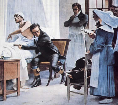
René Théophile Hyacinthe Laënnec and the Stethoscope

By Philip R. Liebson, MD, Rush University, Chicago, Illinois, United States

What constitutes a high-tech instrument? Obviously, in the field of medicine, one that has been developed to improve evaluation of a given condition and lead to a more specific diagnosis. In the early 19th century, there was little that could be considered high-tech in medicine in regard to instrumentation. The physician’s primary means of examining the chest were observation, palpation, and percussion. Actually, percussion of the chest was a novelty of the late 18th century, first developed by Leopold Auenbrugger, an Austrian physician; however, by the end of the century, it had fallen out of favor, except where it had been reintroduced by Nicolas Corvisart, Napoleon’s physician.

It would be expected that a willing student of Corvisart might make inroads into the examination of the chest, especially one who became fascinated with diseases of the chest after seeing his mother die of pulmonary tuberculosis at age six. Thus we introduce René Laënnec (1781-1826). Laënnec, like diagnosticians of the 19th century, was an astute observer. At the time, physicians “listened” to the chest by placing a handkerchief on the chest and the touching the ear to the handkerchief. Do not sneer at this method: as late as the 1920s and 1930s, one of the best cardiology diagnosticians, Lewis Atterbury Conner of Cornell Medical School, used this technique when he became so hard of hearing he could no longer use a stethoscope.

Laënnec, who had studied at the École de Medicin in Paris, had been taught the lost art of percussion by Corvisart. However, he found the use of auscultation with the aid of a handkerchief to be an awkward and frustrating experience, especially when examining obese women. Laënnec, being an observant fellow, noticed children playing with long hollow sticks and when the ear was placed at one end of the stick and a pin scratched at the other end the sound was amplified. Laënnec tried this by using rolled up paper, but also found this somewhat awkward. He was somewhat into carpentry and built a hollow wooden cylinder, 25 cm in length and 2.5 cm in diameter. Thus, a high-tech instrument was borne in the stethoscope. This he accomplished in 1816, while Napoleon, after a quick sojourn at Elba, was now permanently residing in St. Helena. Even though the word “revolution” was out of favor at that time, there is no other term that adequately conveys the import of this discovery; indeed, it had a longer-lasting impact than many of the ideas thought up by the men of the tumbrils several decades before.

Laënnec coined the term “stethoscope” from the Greek words for “chest” and “observer.” He also coined the term “mediate auscultation,” in that the instrument amplified sounds from the chest. His work included descriptions of auscultatory findings in various conditions previously difficult to discern, and introduced the terms such as râles, crepitance, egophony, and rhonchi. Initially, when evaluating heart sounds, he considered that S1 was produced by ventricular systole and S2 to atrial contraction rather than due to closing of the A-V valves and the valves to the great vessels, respectively. His landmark work, published in 1819, was entitled De l’Auscultation Médiate ou Traité du Diagnostic des Maladies des Poumons et du Coeur (On Mediate Auscultation or Treatise on the Diagnosis of the Diseases of the Lungs and Heart).

Let’s step back a minute. In order to fully appreciate Laënnec’s career, one must better understand the times in which he lived. While he was living with his famous uncle, Guillaume-François Laënnec, professor of medicine at the University of Nantes, the French Revolution began. This in no way affected René, who in his pre-teens at the time. He enrolled at the Collège d’Oratorie and studied liberal arts, including religion, political science (one might wonder how the ongoing revolution affected the teaching of this subject), grammar, geography, Latin, verse, and prose. Peaceful life or not, there was a guillotine in Nantes stationed in a square just outside his home, and he had seen several isolated heads produced by that new state-of-the-art procedure invented by Dr. Guillotine to bring death on rapidly in deference to the suffering of the victim. I suppose you could say that it was the precursor of the microtome.

Laënnec began the study of medicine at the age of 14 at his uncle’s hospital in Nantes when the revolution was at its height, and in 1799, along with Napoleon, entered Paris to formally study medicine under Corvisart. I leave it to the reader to decide which one of the two had the greater influence on the 21st century in regard to the individual patient.

His first published work in 1802 was a report of a case of mitral valve calcification with left ventricular dilatation evaluated at autopsy. Just after he received his doctorate, he lectured on tuberculosis. At the time, involvement of the lung was termed phthisis, and he became a savior of lisping physicians by indicating that it was pulmonary tuberculosis. Following this, he spent five years lecturing on pathologic anatomy and doing some private practice. He was also an editor of the Journal de Medicine from 1805 to 1808, between the ages of 24 and 27.

After 1819, when he had published his great work on auscultation, one would have thought that this would incite a revolution in the medical field in France. Wrong! Unfortunately, there was no Robespierre or Marat in medicine to foster his ideas and indifference was the attitude among his colleagues. However, the rest of Europe was enthusiastic, and physicians came from all over the continent to the Necker Hospital to see Laënnec demonstrate his new instrument. There is no information available on whether they received CME credits or had drug reps in the adjoining rooms. His book, of course, was a sellout outside of France. Finally, his French colleagues accepted his talents and he was made a professor of medicine at the Collège de France in 1822 at age 41 and a member of the Academy of Medicine. Several years later he was made a knight of the Legion of Honor. In 1826, still only 45 years of age, he developed a cold followed by a throat infection and chest pains. He asked his nephew to listen to his chest and describe what he heard. Laënnec interpreted the findings as cavitary tuberculosis. This caused his death. He died before Chopin developed full-fledged tuberculosis, but it would be interesting to surmise what Laënnec could have accomplished had he been the composer’s physician. At least we would have had significantly more knowledge of the extent of his condition.

In his will, Laënnec bequeathed to his nephew all his scientific papers and his stethoscope, which he indicated was “the best part of my legacy.” In 1867 money was raised in Europe for a monument to him at the Paris Exposition the following year. The modern binaural stethoscope was invented in the 1850s and the rest, as they say, is history. Now every practicing doctor, except psychiatrists, pathologists, and perhaps some dermatologists, carries around a stethoscope, and, despite the availability of echocardiography, chest radiography, CT scanning, and MRI, may occasionally use it. It is unlikely that any of them carry around Napoleon’s picture.

References
1. Willius FA, Keys TE. Classics of Cardiology, Volume One. René Théophile Hyacinthe Laënnec pp. 323-382. Dover Publications, New York. 1941
2. Roguin A. René Theophile Hyacinthe Laënnec (1781-1826): The man behind the stenoscope. Clinical Medicine and Research 2006;4:230-235.
3. Fayssoil A. René Laennec (1781-1826) and the invention of the stethoscope. Am J Cardiol 2009; 743-744
4. Scherer JR. Before cardiac MRI: René Laennec (1781-1826) and the invention of the stethoscope. Cardiology Journal 2007;14:518-519.
5. Tan SY, Yeow ME. Medicine in Stamps René Laennec (1781-1826): Inventor of the stethoscope. Singapore Med J 2005;46: 106-107.

PHILIP R. LIEBSON, MD, graduated from Columbia University and the State University of New York Downstate Medical Center. He received his cardiology training at Bellevue Hospital, New York and the New York Hospital Cornell Medical Center, where he also served as faculty for several years. A professor of medicine and preventive medicine, he has been on the faculty of Rush Medical College and Rush University Medical Center since 1972 and holds the McMullan-Eybel Chair of Excellence in Clinical Cardiology.

Image: Laennec examines a consumptive patient with a stethoscope in front of his students at the Necker Hospital, 1816. Théobald Chartran

From: hektoeninternational.org

Sunday, December 6, 2015

A Civil War Medic’s Knapsack

By Chuck Franson, Registrar, AMEDD Museum, Number 3, Summer 2013

At the beginning of the Civil War, each regimental surgeon was outfitted with equipment and supplies for his regiment, including medicines, stores, instruments, and dressings, in quantities regulated by the Standard Supply Table for Field Service. In the field he was accompanied by a hospital orderly, who carried a knapsack containing a limited supply of anesthetics, styptics, stimulants, anodynes, and material for primary dressings.

An army board had recommended this hospital knapsack for adoption in 1859. It was made of light wood or wicker, and was covered with canvas or enameled cloth; it weighed about 18 pounds when full. This knapsack was in general use in the first year of the war, but it was difficult to keep items in order. In 1862 it was replaced with the “Regulation Hospital Knapsack of 1862”, which featured a better arrangement of supplies and medications. The new pattern was 16 inches high, 12½ inches wide, and 6 inches deep; the contents were packed in 3 drawers, which were more accessible than in the old style and less liable to become disarranged or broken.

The contents of the knapsack were:
One piece of white wax, 8 oz. simple cerate, 12 oz. chloroform, 5 yds. adhesive plaster, 2 yds. isinglass plaster, 1 oz. persulphate of iron, 100 compound cathartic pills, 150 blue mass pills, 150 opium pills, 100 opium and camphor pills, 150 quinine pills, 8 oz. aromatic spirit of ammonia, 16 oz. brandy, 4 oz. laudanum, 10 bandages, 10 binder's boards, 4 oz. charpie, 2 medicine glasses, 1 (spirit) lamp, 12 oz. lint, 1 box matches, 1 paper of pins, 1 spool of surgeons' silk, 4 pieces of sponge, 4 (Dunton's) field tourniquets. 2 spiral tourniquets, 1 piece of tape, 1 spool of lead wire, 1 spool of silver wire, and 1 spatula.

Weight when packed was nearly 20 pounds. Despite its convenience and general adaptability it was too unwieldy to be carried by the surgeon himself, and was liable to be lost in action when left to anyone else.

It was replaced in early 1863 by a field case or “Surgeon’s Companion” designed by Medical Inspector R. H. Coolidge, similar to a British model, to be carried by the surgeon himself if necessary.

This one contains:
Top Drawer
1 Scarificator
2 Tourniquets
2 rolls of cotton tape ½” wide
8 Wooden slates (splints)
1 Container Plaster
1 Roll of Adhesive Plaster w/o container
1 Bloodletting knife

Middle Drawer
33 Rolls Bandages
1 Pewter container Pilcathart Co.
1 Pewter container Pil Opit et camph
1 Pewter Container Chloroformum
1 Scarifacator in box

Bottom Drawer
3 Dunton’s Tourniquets
Pewter Containers: 2 Pil Hydrarg; Pil
Quiniae Sulph; Pil Opit; Sp Ammon
Ar: Chloroformum; 2 Ceratum Simp;
Opit; Pil Opit et Camph; Sp Vini Gale

From: history.amedd.army.mil/newsletters

Collidge Field Case or Surgeon's Companion

From: medicalantiques.com

(The following are the personal edited research notes of Michael Echols, the source of which may or may not be completely documented)

In the early part of 1863 Medical Inspector R. H. Coolidge, U. S. A.. arranged a field case or companion  (FIG. 1) to take the place of the knapsack. It was based on the plan of the one used in the British service, and was intended to be carried by the Surgeon himself, if necessary.   The Surgeon's Field Companion is one variation used during the Civil War.

The "companion" is a leather case 13 inches long, 6 inches wide, and 7½ inches deep; it is supported by a strap passing over the shoulder, and is provided with a waist strap to steady it when carried.

The hospital medicine chest, mess-chest, and bulky hospital supplies were transported in wagons of the supply train and were often inaccessible when required. To obviate this, the contents of the knapsack were:

One piece of white wax, 8 oz. simple cerate, 12 oz. chloroform, 5 yds. adhesive plaster, 2 yds. isinglass plaster, 1 oz. persulphate of iron, 100 compound cathartic pills, 150 blue mass pills, 150 opium pills, 100 opium and camphor pills, 150 quinine pills, 8 oz. aromatic spirit of ammonia, 16 oz. brandy, 4 oz. laudanum, 10 bandages, 10 binder's boards, 4 oz. charpie, 2 medicine glasses, 1 (spirit) lamp, 12 oz. lint, 1 box matches, 1 paper of pins, 1 spool of surgeons' silk, 4 pieces of sponge, 4 (Dunton's) field tourniquets. 2 spiral tourniquets, 1 piece of tape, 1 spool of lead wire, 1 spool of silver wire, and 1 spatula.

The contents of the Surgeon's Companion were;

6½ oz. chloroform, 2 oz. fluid extract of ipecacuanha. 2 oz. fluid extract of ginger. 2 oz. solution of persulphate of iron, 24 oz. of whiskey, 2 oz. tincture of opium, 144 compound cathartic pills, 144 colocynth and ipecacuanha pills, 144 sulphate of quinine pills, 144 opium pills, 1 yard isinglass plaster, a medicine cup, scissors, teaspoon, pins, thread, 4 oz. lint, a towel, 2 doz. bandages, muslin, and corks.

At the beginning of the war each regimental Surgeon was furnished with suitable equipment for his regiment for field service, consisting of medicines, stores, instruments, and dressings, in quantities regulated by the Standard Supply Table.

(See Standard Supply Table for Field Service, in Revised Regulations for the Army of the United States, 1861, p. 304.)

In action he was accompanied by a hospital orderly, who carried a knapsack containing a limited supply of anaesthetics, styptics, stimulants, and anodynes, and material for primary dressings. This hospital knapsack had been recommended for adoption by an army board in 1859; it was made of light wood, 18 inches in height, 15 inches wide, and 7½ inches deep, but subsequently wicker-work, covered with canvas or enamelled cloth, was substituted for the wood; its weight when filled was 18 pounds. This knapsack (FIG. 425) was in general use in the first year of the way and served an excellent purpose. In 1862 it was changed for what was known as the new regulation knapsack, in which the arrangement and character of the supplies were modified. The new pattern was 16 inches high, 12½ inches wide, and 6 inches deep; the contents were packed in drawers, which were more accessible than in the old style and less liable to become disarranged or broken.(1) The weight when packed was nearly 20 pounds. (FIG. 2). Not withstanding its convenience and general adaptability it was too heavy and cumbrous to be carried by the Surgeon himself, and, when entrusted to other hands, was liable, in the vicissitudes of battle, to be lost.
     
Apparently  there were four military field medicine carrying cases used by the Union army during the Civil War.  In 1861, a large wicker and tarred-cloth knapsack was the regulation case (Fig. 2).   This was superseded by a heavy wood-framed and leather-covered backpack (Fig. 2) 1862.   In 1863, the Coolidge Field, or Surgeon’s Companion, which was worn on the hip became the regulation case.  There was a final version, also worn on the hip, but it is not shown in the illustrations of the Medical and Surgical History data.

Image: Fig. 2  Hospital Knapsack of Wicker-Work, Covered With Enamelled Cloth.

Image: Fig. 3 Regulation Hospital Knapsack of 1862

(Note: in the drawings, all tins and bottles are shown with corks and not metal screw tops.)

Share

Facebook Twitter Delicious Stumbleupon Favorites